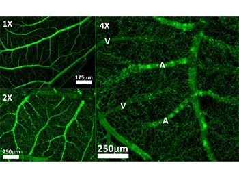
The correct imaging of blood vessels is of crucial importance in the diagnosis and treatment of vascular pathologies, as well as in the conduct of clinical research. However, most methods for studying the evolution of blood vessels are invasive and require injections of toxic products, such as contrast products, fluorophores or radioactive markers, associated with techniques (synchrotron, coherence tomography optics, etc). Researchers have developed a unique algorithm that can extract at very high resolution, in just a few minutes, the vascularization of a flat organ such as the chorioallantoic membrane (CAM) of a chicken or an eye, simply using a single HD camera and the red blood cells themselves as flow tracers. There is no need for expensive imaging devices or specific contrast agents, if the red blood cells can be traced. The algorithm is particularly suitable for microvascular imaging, in animal models such as chickens or rabbits. It may be of interest for research in ophthalmology, oncology on neovascularization and induced ischemia testing. Time-lapse imaging reveals the rich dynamic behavior of vessels in pathological instances and recovery pathways
Imaging - Software - Vasculature Diagnostic - Research
Erganeo se tient à votre écoute.